Varghese Alex, Kiran Vaidhya, Subramaniam Thirunavukkarasu, Chandrasekharan Kesavadas, Ganapathy Krishnamurthi
Journal of Medical Imaging, Vol. 4, Issue 04, 041311, (December 2017) https://doi.org/10.1117/1.JMI.4.4.041311
TOPICS: Neurons, Brain mapping, Neodymium, Tumors, Image segmentation, Brain, Denoising, Neuroimaging, Magnetic resonance imaging, Computer programming
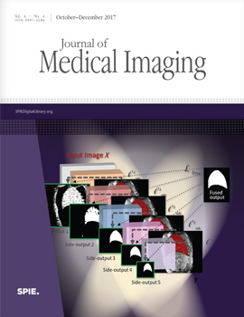
The work explores the use of denoising autoencoders (DAEs) for brain lesion detection, segmentation, and false-positive reduction. Stacked denoising autoencoders (SDAEs) were pretrained using a large number of unlabeled patient volumes and fine-tuned with patches drawn from a limited number of patients ( n=20, 40, 65). The results show negligible loss in performance even when SDAE was fine-tuned using 20 labeled patients. Low grade glioma (LGG) segmentation was achieved using a transfer learning approach in which a network pretrained with high grade glioma data was fine-tuned using LGG image patches. The networks were also shown to generalize well and provide good segmentation on unseen BraTS 2013 and BraTS 2015 test data. The manuscript also includes the use of a single layer DAE, referred to as novelty detector (ND). ND was trained to accurately reconstruct nonlesion patches. The reconstruction error maps of test data were used to localize lesions. The error maps were shown to assign unique error distributions to various constituents of the glioma, enabling localization. The ND learns the nonlesion brain accurately as it was also shown to provide good segmentation performance on ischemic brain lesions in images from a different database.


 Receive Email Alerts